In part 2 I would like to deal with the muscular problems that arise from the hoof position: long toe and underrun heels.
In the previous part I referred to the problems and damages of the joint and ligament structures at the distal part of the limb that may occure swith long toe and underrun heels.
I’m trying to explain this as simply as possible, so please bear with me if I leave out a few facts you may have noticed. Certainly this article is not complete and perhaps there are a few examples that are somewhat different.
What are the digital flexor tendons for?
First of all, of course, to bend the carpal joint, fetlock joint, pastern joint (superficial toe flexor) and hoof joint (deep toe flexor).
Another very important task of the flexor tendons is to absorb the momentum of the forward movement and the weight of the horse’s body, which rests with 56% on the front limb. Therefore the flexor tendons belong to the so-called energy storing tendons. They can absorb high loads in relatively short consecutive repetitions without being damaged if they relax completely and are sufficiently supplied with blood afterwards.
Owners often tell that their horse stepped into a hole, got scared and slipped in a tight turn or something similar and injured the suspensory ligament or the deep/superficial flexor tendon.
So I have a question: horses in the wild fleeing from a predator over uneven terrain, how often do they get into a hole unexpectedly or make tight turns without hurting themselves? So is it the turn or the uneveness of the ground that causes the tendon injury? With an old horse maybe yes, otherwise certainly not.
This means that something must happen to the digital flexor tendons that limits the extensibility of these tendons and the support ligaments (proximal support ligament above the carpus belongs to the superficial flexor tendon, distal support ligament directly below the carpus belongs to the deep flexor tendon).
And here we come to the third task of the digital flexor tendons. Like some other tendons and muscles of the anterior limb, they belong to the passive standing apparatus of the anterior limb.
How does this passive standing apparatus work?
You can certainly write a whole chapter in a veterinary scinece book about this in its subtleties (if you want you can read everything in such a book). But simply put there is a muscle tract on the dorsal side of the frontleg, which takes over the holding function on the front leg against unintended flexion (almost all parts of the extensor muscles). And there is another muscle tract on the palmar side of the leg against nintended extension (almost all flexors), so the horse is able to keep standing without tiring its muscles.
That’s why these muscles have a large proportion of special fibres (type 1) which are very enduring and hardly tiring. This enables them to perform a permanent holding function. Otherwise, muscles and their health depend on continuous contraction and relaxation. Only in this way are they sufficiently supplied with blood and metabolic products transported away.
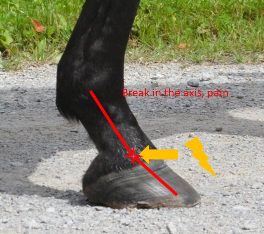
The change in the angle of the coffin bone (long toe, underrun heels > increased extension, angle of coffin bone to ground > 6°, pontentially negative) results in a permanent increase in traction on the digital flexor tendons (usually not on the deep flexor tendon, because the coffin joint is still standing under the centre of gravity in flexion). Hence the still possible hyperextension as you can see in the picture. Hence the frequency distribution of the injury with 87% superficial flexor tendon and only 13% deep flexor tendon.
Joints such as the hoof joints , pastern joints, fetlock joints and carpal joints are thus permanently in a tendency to overstretching. And joints also have receptors that regulate the tension of the muscles. To protect themself against injuries.
The nervous system reacts to this overstretching of tendons, but above all to that of the joints. Once with a reduction in the tone of the extensor muscles, which is why the flexors of the passive standing apparatus contract more strongly than their opponents the extensors. In addition and above all, passive hyperextension strengthens the tone in the flexor muscles, combined with reduced blood circulation in the muscle, which further limits its elasticity.
From this you can deduce that the muscle of the superficial digital flexor (which is more affected than the deep flexor tendon because of the stretching of the fetlock joint due to the position of the hoof)
1. is contracted
Tension acting on the flexor tendons has a stronger effect on the tendons, as the muscle is less elastic.
2. poorly supplied with blood
Tears of muscle fibers and other damages will happen more often
And not only the muscle of the superficial digital flexor tendon is less supplied with blood. The blood circulation of the tendon itself is also disturbed.
A flexor of the carpal joint that is particularly noticeable in the chain of the passive standing apparatus is the M. extensor carpi ulnaris. The muscle is called extensor, but in the horse it is in fact a flexor and reaches from the elbow to the os pisiforme. He is largely responsible for the visible flection in the phase of extension of the frontleg.
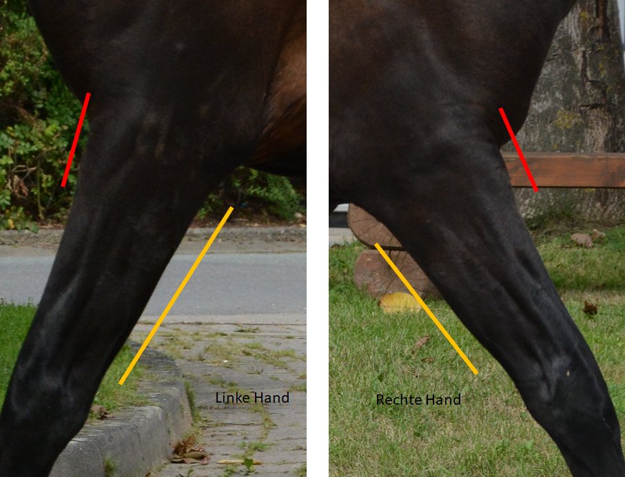
The Extensor carpi radialis (flexor) on the right hand cannot be stretched as much as that of the left hand, recognizable by the
small muscle belly above the yellow line. The muscle of the left hand is more stretched (almost straight line on the back of the
forearm). In addition, one sees that the carpal joint extensor (marked by red line) on the right hand must contract much more
strongly than that of the left side (muscle belly protrudes more clearly).
The increased tone of the M. flexor carpi ulnaris pulls the os pisiforme in the direction of flexion even at the moment of carpal joint extension. The pressure of the os pisiforme and the flexor cuff around the carpal joint restricts the blood supply to the digital flexor tendons themselves and thus prevents a regeneration of the digital flexor tendons in the short term during the relaxation phase. In the long term, even the structure of the tendons and its molecular structure will change, allowing it to suffer even more easily micro-cracks and worse injuries.
Furthermore, the increased tension in the superficial digital flexor tendon leads to a minimal flexion in the crown joint, thus to instability of the crown joint.
If we follow the problem further upwards into the front limbs, we often find that horses with long toes and underrun heels also have problems with stretching the shoulder joint. Their forward move especially in the opposite frontleg looks flat, it’s not a wideranged efficient movement. This explains because the biceps (also part of the passive standing apparatus) is also needed to prevent overstretching of the carpal joint. However, since the biceps also flexes the shoulder, the necessarily increased tension in the biceps leads to flexion in the shoulder.
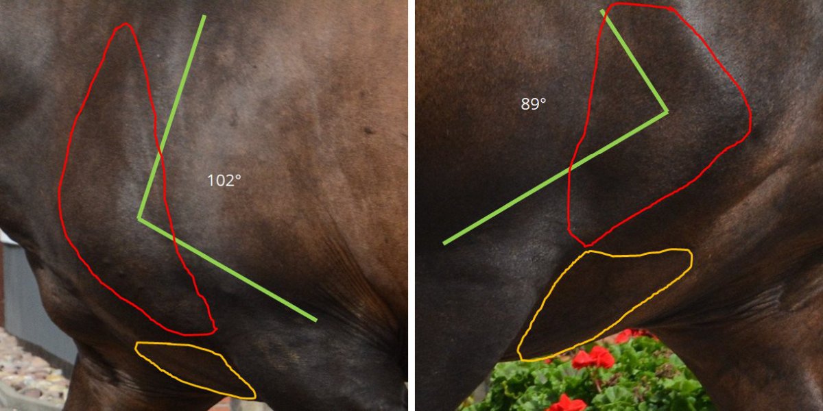
At 102°, the left shoulder is significantly wider than the right shoulder, which remains in a bending position at less than 90°. The red field shows the biceps muscles and the infra- and supraspinatus muscles (all flexors of the shoulder joint at an angle below 90° and parts of the passive holding apparatus). On the right shoulder you can clearly see that this area does not loosen – as it would be necessary in the extension of the shoulder, but that due muscles have to stretch under tension. It looks as if the horse has less muscles at this point and the bone structures become more prominent. You can also see in the yellow field, that the M.pectoralis transversus is more toned with the right leg, pulling the leg medial to compensate the functional shortening of the right frontleg.
If one then looks at the front limb altogether (flexion shoulder, flexion carpal joint, overstretching toe) the affected side is functionally shorter than the other forehand.
This leads to rotation in the 6th, 7th cervical vertebra, 1st thoracic vertebra and 1st rib (because the shoulder limb is pulled towards the body to compensate for the difference in length) and, in the case of sudden uncontrolled movements, to a blockage of the affected vertebrae. This means that the horse is no longer able to bend these vertebrae and thus the neck in the opposite direction (to the left side).
The base narrow position of the affected leg will lead to further problems with the hoof, espacially concerning the latero-medial equilibrium of the hoof. Often problems with this equilibrium are based on failures of the dorso-palmar equilibrium and are wrongly seen as stand alone issue.
But the whole story doesn’t end there. Of course, restrictions in the cervical spine lead to compensatory movements across the entire spine from the head joint to the sacroiliac joint. Blockages in the head joint lead to problems in the jaw joint and the teeth and blockages in the sacroiliac joint can lead to problems in the knee and ankle joint.
So you see that
1. a lameness in the hind leg can be caused by the front leg
so it makes no sense to treat a horse only where I see a conspicuous lameness.
2. very many problems in the area of the cervical spine and the sacroiliac joint come out of the legs
3. dental problems often originate from the neck / chest
4. it’s worth to pay a good farrier a little more so he takes his time for you and your horse
a correct shoeing with calculation of the rolling point, rocking and forging of iron, gait evaluation
etc. simply takes at least 2 hours and a capable farrier should be paid fairly, a good job never comes cheap